

 0
0 0
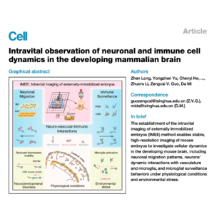
0Researchers have developed a novel intravital imaging method called IMEE (externally immobilized embryos) to observe cellular dynamics in the developing mouse brain with unprecedented clarity. This technique allows for long-term, deep-tissue observation of living embryos while maintaining a stable intrauterine environment and normal blood circulation. By utilizing this method, the study characterizes the migration patterns of neurons and the behavior of immune cells like microglia under both healthy conditions and environmental stress. The findings reveal how cells adapt their movements to local environments, specifically highlighting interactions between neurons, blood vessels, and the immune system. Furthermore, the researchers utilized this tool to identify migratory defects in models of neurodevelopmental disorders. Ultimately, this platform provides a powerful means to study mammalian brain development and real-time cellular responses in vivo.
References:
- Long Z, Yu Y, He C, et al. Intravital observation of neuronal and immune cell dynamics in the developing mammalian brain[J]. Cell, 2025.
