

 1
1 0
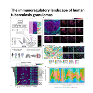
0This research utilizes multiplexed ion beam imaging (MIBI-TOF) to construct a spatial atlas of the human tuberculosis (TB) granuloma, revealing how immune cell organization dictates disease progression. By analyzing archival tissues, the authors identified eight distinct microenvironments where specific myeloid and lymphoid cells interact, highlighting a specialized myeloid core characterized by immunosuppressive proteins like PD-L1 and IDO1. Interestingly, while these regulatory markers are prevalent in active lesions, the typical T cell exhaustion seen in cancer is absent, suggesting a unique myeloid-mediated suppression mechanism. To validate these findings, the study integrated transcriptomic data from over 1,500 patients, proving that elevated PD-L1 levels in the blood can predict the transition from latent to active TB. Ultimately, this work provides a framework for understanding why some infections persist and suggests that host-directed therapies must account for the local spatial dynamics of the immune response.
References:
- McCaffrey, E.F., Donato, M., Keren, L. et al. The immunoregulatory landscape of human tuberculosis granulomas. Nat Immunol 23, 318–329 (2022). doi.org
