

 0
0 0
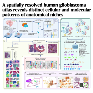
0This study presents a comprehensive, high-resolution atlas of human glioblastoma, documenting the complex cellular and molecular organization within the tumor's distinct anatomical regions. By integrating spatial transcriptomics, single-cell RNA sequencing, and protein measurements, the researchers mapped 56 distinct cell states across more than 115,000 spatial data points. They identified previously uncharacterized vascular, stromal, and immune cell populations, including a specific oligodendrocyte subtype found in the tumor core that is linked to poor clinical outcomes. The authors also defined ten anatomical feature niches, such as the Immune-Glial Niche (IGN) and microvascular proliferation, to better understand how the microenvironment shapes tumor behavior. This multi-modal resource is publicly accessible and provides a detailed framework for identifying therapeutically actionable vulnerabilities in aggressive brain cancer.
References:
- Shah N, Park H J, Sonpatki P, et al. A spatially resolved human glioblastoma atlas reveals distinct cellular and molecular patterns of anatomical niches[J]. 2024.
