

 0
0 0
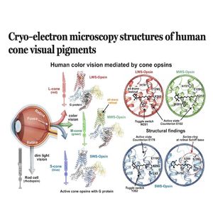
0This research article details the first high-resolution cryo-electron microscopy structures of the three human cone opsins responsible for color vision: long-, middle-, and short-wavelength-sensitive pigments. By capturing these proteins in their active states complexed with a G protein, researchers identified unique molecular features that distinguish color vision from the dim-light vision mediated by rhodopsin. Key findings include a specialized "serine ring" in blue-sensitive opsins and a different counterion switch mechanism in red and green pigments, which together facilitate spectral tuning and rapid signal decay. The study further clarifies the molecular basis of color vision deficiencies by mapping pathogenic mutations onto these new structural models. Ultimately, these results provide a foundational framework for understanding the biophysical mechanisms of human phototransduction and the origins of various retinal disorders.
References:
Peng Q, Li J, Jiang H, et al. Cryo–electron microscopy structures of human cone visual pigments[J]. Science, 2026, 392(6805): eadz8141.
